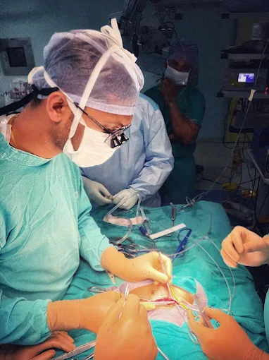
Treatment

Urological Cancer

Urological Cancer

Urological Cancers affect the organs of the urinary tract and the male reproductive system, including the kidneys, bladder, prostate, and testicles.
These cancers are among the most common, especially prostate cancer in men.
Early signs may include blood in the urine, pain during urination, or lower back pain.
Regular screening is vital for early detection and effective treatment.
Advanced diagnostic tools help in accurate assessment and staging of the disease.
Treatment options vary from surgery and radiation to chemotherapy and immunotherapy.
Consulting a urology specialist can significantly improve patient outcomes and recovery.

Kidney Cancer

Kidney cancer, also known as renal cancer, is a type of cancer that starts in the kidneys β€” the two bean-shaped organs located behind your abdominal organs, with one kidney on each side of your spine.

Types of Kidney Cancer

The most common types include:

Renal Cell Carcinoma (RCC):

The most common form (~90% of adult kidney cancers).

Usually develops in the lining of the kidney's tubules.

Transitional Cell Carcinoma:

Arises in the renal pelvis (where the kidney meets the ureter).

Similar to bladder cancer.

Wilms Tumor:

Most common in children, especially under age 5.

Renal Sarcoma:

Rare type that begins in the connective tissue of the kidney.

Symptoms

Kidney cancer often doesn’t cause symptoms in early stages. As it progresses, common symptoms may include:

Blood in the urine (hematuria)

Persistent pain in the back or side

A mass or lump in the abdomen

Fatigue

Unexplained weight loss

Fever that isn’t caused by infection

Anemia (low red blood cell count)

Risk Factors

Smoking
Obesity
High blood pressure
Family history of kidney cancer
Certain inherited conditions (e.g., von Hippel-Lindau disease)
Long-term dialysis

Diagnosis

Imaging tests: CT scan, MRI, ultrasound
Urine and blood tests
Biopsy (in some cases)

Treatment Options

Treatment depends on the type, stage, and overall health:

Surgery:

Partial nephrectomy: Removal of the tumor only.

Radical nephrectomy: Removal of the entire kidney.

Targeted therapy:

Drugs that block specific pathways cancer cells use to grow.

Immunotherapy:

Boosts the body’s immune response against cancer.

Radiation therapy:

Less commonly used, typically for pain management or if surgery isn't an option.

Active surveillance:

In slow-growing tumors or for patients unable to undergo aggressive treatment.

Prognosis

The outlook depends on the stage at diagnosis and the overall health of the patient.

Early-stage kidney cancer often has a good prognosis if surgically removed.

Laparoscopic Radical Nephrectomy is a minimally invasive surgical procedure used to remove an entire kidney, typically due to kidney cancer or other serious kidney diseases.

Key Points:

πŸ” What does "Radical Nephrectomy" mean?

Radical = complete removal

Nephrectomy = removal of the kidney

So, a radical nephrectomy involves removing:

The entire kidney

The adrenal gland (in some cases)

Surrounding fatty tissue

Sometimes nearby lymph nodes

πŸ”§ What does "Laparoscopic" mean?

It refers to a minimally invasive technique using small incisions and a camera (laparoscope).

The surgeon uses special instruments inserted through these incisions to perform the surgery.

Compared to open surgery, it usually results in:

Less pain

Smaller scars

Shorter hospital stay

Faster recovery

Indications:

Renal cell carcinoma (kidney cancer)

Large or complex benign tumors

Procedure Overview:

General anaesthesia is administered.

3–5 small incisions are made in the abdomen.

A laparoscope (camera) is inserted to guide the surgery.

The kidney and surrounding structures are detached and removed, usually through one slightly larger incision.

The incisions are closed.

Adrenal Cancer

Adrenocortical cancer (ACC) β€” also known as adrenal cortical carcinoma β€” is a rare and aggressive cancer that originates in the outer layer (cortex) of the adrenal glands. The adrenal glands sit atop the kidneys and are responsible for producing hormones such as cortisol, aldosterone, and androgens (sex hormones).

πŸ” Overview

Rarity: ACC is very rare, affecting about 1–2 people per million each year.

Aggressiveness: Often diagnosed at an advanced stage and may spread (metastasize) to other organs.

Age groups: Can occur at any age but has two peak incidences β€” in children under 5 and adults in their 40s–50s.

⚠️ Symptoms

Symptoms depend on whether the tumor is functioning (hormone-producing) or non-functioning:

Functioning Tumors (most ACCs)

Produce excess hormones, causing symptoms like:

Cushing’s syndrome (excess cortisol):

Weight gain (especially in the face and abdomen)

Purple stretch marks

Muscle weakness

High blood pressure and blood sugar

Virilization or feminization (excess sex hormones):

In women: facial hair, deepening voice, irregular periods

In men: breast enlargement, decreased libido

Hyperaldosteronism (excess aldosterone):

High blood pressure

Low potassium

Non-Functioning Tumors

Flank or abdominal pain

Palpable abdominal mass

Unexplained weight loss

Fatigue

πŸ§ͺ Diagnosis

Imaging: CT or MRI scan of the abdomen to assess the adrenal mass.

Hormone testing: To detect overproduction of cortisol, aldosterone, or androgens.

Biopsy: Rarely done due to risk of spreading cancer cells β€” diagnosis usually confirmed after surgical removal.

PET scan or bone scan: To check for metastasis.

🧬 Causes and Risk Factors

Most ACCs occur sporadically, but some are linked to genetic syndromes, including:

Li-Fraumeni syndrome

Beckwith-Wiedemann syndrome

Multiple endocrine neoplasia type 1 (MEN1)

Familial adenomatous polyposis (FAP)

πŸ› οΈ Treatment

Treatment depends on stage and whether the tumor is producing hormones:

Surgery (Mainstay treatment):

Complete surgical removal (adrenalectomy) is the goal, often with surrounding tissue.

Surgery may be curative in early stages.

Mitotane (Lysodren):

An adrenal-specific drug used to destroy adrenal tissue.

Often used after surgery to reduce recurrence or for metastatic disease.

Chemotherapy:

Regimens may include drugs like etoposide, doxorubicin, and cisplatin (EDP) plus mitotane.

Used for advanced or recurrent disease.

Radiation therapy:

May be used after surgery or for symptom control in metastatic disease.

Targeted and Immunotherapy:

Being investigated in clinical trials, but not yet standard treatments.

πŸ“Š Prognosis

Early-stage ACC (Stage I or II): Surgery may be curative.

Advanced-stage ACC (Stage III or IV): Prognosis is poorer, especially if metastases are present.

5-year survival rates:

Stage I: ~80%

Stage IV: < 20%

Laparoscopic Adrenalectomy is a minimally invasive surgical procedure to remove one or both adrenal glands, which sit above the kidneys and produce important hormones like cortisol, aldosterone, and adrenaline.

This procedure is done using a laparoscope (a thin camera) and specialized instruments inserted through small incisions in the abdomen or flank.

🩺 Why is Laparoscopic Adrenalectomy Done?

It’s typically used to treat:

Adrenal tumors, including:

Functioning (hormone-producing) tumors, such as:

Pheochromocytoma – produces excess adrenaline

Aldosteronoma (Conn’s syndrome) – overproduces aldosterone

Cortisol-producing adenoma (Cushing's syndrome)

Non-functioning adrenal masses (usually >4–6 cm or growing)

Adrenocortical carcinoma (in selected cases)

Metastasis from other cancers

πŸ”§ How Is It Performed?

General anesthesia is administered.

The surgeon makes 3–5 small incisions in the abdomen or flank.

A laparoscope and surgical tools are inserted.

The adrenal gland is carefully separated from surrounding structures (e.g., kidney, major blood vessels).

The gland is placed in a retrieval bag and removed through one of the incisions.

Incisions are closed; a drain may be placed temporarily.

🧭 Approaches:

Transabdominal (most common): through the front or side of the abdomen

Posterior retroperitoneoscopic: through the back, avoiding abdominal organs (good for small tumors)

βœ… Advantages of Laparoscopic Over Open Adrenalectomy:

Smaller incisions, less scarring

Less pain after surgery

Shorter hospital stay (1–3 days)

Faster return to normal activities

Lower risk of infection and complications

🧬 One or Both Glands?

Unilateral adrenalectomy: Most common (for tumors on one side)

Bilateral adrenalectomy: Rare; needed in some cases of Cushing’s disease, bilateral tumors, or genetic syndromes

πŸ₯ Recovery:

Most patients go home in 1–3 days

Full recovery in 2–4 weeks

Hormonal monitoring continues post-op

If both glands are removed, lifelong hormone replacement is required

Cancer of Ureters

UTUC stands for Upper Tract Urothelial Carcinoma. It's a relatively rare type of urothelial cancer that occurs in the lining of the upper urinary tract, specifically in the:

Renal pelvis (part of the kidney that collects urine)

Ureters (tubes that carry urine from the kidneys to the bladder)

πŸ” Overview

UTUC accounts for 5–10% of all urothelial cancers.

More common in older adults, typically in their 70s.

It shares many characteristics with bladder cancer, which also originates from the urothelium, but UTUC tends to be more aggressive and difficult to detect early.

⚠️ Symptoms

Often nonspecific or silent in early stages:

Hematuria (blood in the urine) – most common symptom
Flank pain
Urinary tract infections
Unexplained weight loss or fatigue (in advanced cases)

πŸ§ͺ Diagnosis

Urinalysis and cytology:

Detect blood and abnormal cells in urine.

Imaging:

CT urogram (gold standard): Provides detailed images of the kidneys, ureters, and bladder.

MRI or ultrasound may also be used.

Ureteroscopy:

A scope is inserted into the urinary tract to visualize the tumor and obtain a biopsy.

🧬 Risk Factors

Smoking (most significant)

Occupational exposure to chemicals (e.g., aromatic amines, used in dye and chemical industries)

Chronic inflammation or urinary tract infections

Lynch syndrome (hereditary cancer syndrome)

History of bladder cancer

πŸ“Š Staging

UTUC is staged similarly to other urothelial cancers:

Non-muscle-invasive (low-grade, localized) – confined to the urothelium or lamina propria

Muscle-invasive (high-grade, invasive) – penetrates deeper layers, possibly into surrounding organs or lymph nodes

πŸ› οΈ Treatment

Treatment is based on tumor location, grade, and stage:

1. Low-grade, non-invasive UTUC:

Endoscopic ablation or resection (using laser or electrocautery via ureteroscopy)

Surveillance with regular imaging and ureteroscopy

2. High-grade or invasive UTUC:

Nephroureterectomy (removal of the kidney, ureter, and part of the bladder)

Lymph node dissection

Adjuvant chemotherapy (typically cisplatin-based) may be given before or after surgery

3. Intracavitary therapy (e.g., BCG or mitomycin) is under investigation for low-grade UTUC but less effective than in bladder cancer due to anatomy and drug delivery challenges.

🎯 Prognosis

Low-grade UTUC: Often curable with endoscopic or surgical treatment

High-grade/invasive UTUC: Has a higher risk of recurrence and metastasis

5-year survival rates depend on stage and grade:

Localized: ~60–90%

Regional spread: ~30–50%

Distant metastasis: ~5–10%

Laparoscopic Radical Nephroureterectomy is a minimally invasive surgical procedure in which the entire kidney, ureter, and a portion of the bladder where the ureter inserts (called the bladder cuff) are removed. This surgery is primarily performed to treat upper urinary tract urothelial carcinoma (UTUC) β€” a type of cancer that affects the lining of the kidney and ureter.

🩺 What is it for?

The procedure is typically indicated for:

Urothelial carcinoma of the kidney or ureter

High-grade or invasive tumors

Tumors that are not suitable for kidney-sparing treatments

🧬 What is Urothelial Carcinoma?

A cancer arising from the urothelium β€” the lining of the renal pelvis, ureter, and bladder

Similar to bladder cancer but located higher in the urinary tract

Often requires complete removal of the kidney and ureter to prevent recurrence

πŸ”§ How is Laparoscopic Radical Nephroureterectomy Performed?

General anesthesia is administered.

Multiple small incisions are made in the abdomen or flank.

A laparoscope (camera) and instruments are inserted.

The surgeon removes:

The entire kidney

The entire ureter

The bladder cuff (the part of the bladder where the ureter connects)

Lymph nodes may be removed if cancer spread is suspected.

A specimen retrieval bag is used to remove the organs through a slightly larger incision.

πŸ₯ Recovery:

Hospital stay: 2–4 days

Catheter: Often left in place for 1–2 weeks

Return to normal activities: 2–4 weeks

Full recovery: 4–6 weeks

βœ… Advantages of Laparoscopic Approach:

Less blood loss

Smaller incisions and better cosmetic results

Shorter hospital stay and faster recovery

Comparable cancer control to open surgery

πŸ†š Compared to Other Procedures:

Procedure
Scope
Used For
Laparoscopic Radical Nephroureterectomy Kidney + ureter + bladder cuff UTUC (high-grade)
Laparoscopic Nephrectomy Kidney only Kidney cancer (renal cell carcinoma)
Segmental Ureterectomy Part of ureter Low-grade, localized ureteral tumors
Endoscopic Resection Tumor only Select low-risk, small tumors

Bladder Cancer

Bladder cancer is a common type of cancer that begins in the cells lining the inside of the bladder, a hollow organ in the pelvis that stores urine.

πŸ” Overview

Most common type: Urothelial carcinoma (transitional cell carcinoma)

Arises from the urothelial cells lining the bladder

Can also affect the ureters and renal pelvis (like in UTUC)

Other rare types:

Squamous cell carcinoma

Adenocarcinoma

Small cell carcinoma

⚠️ Symptoms

Bladder cancer often presents with:

Hematuria (blood in the urine) β€” most common early sign

May be visible (gross hematuria) or microscopic

Frequent urination

Pain or burning during urination

Urgency to urinate

Pelvic or back pain (in advanced disease)

🧬 Risk Factors

Smoking (major risk factor β€” causes >50% of cases)

Chemical exposures (e.g., dyes, rubber, leather industries)

Chronic bladder irritation (from infections, catheters)

Previous radiation or chemotherapy (e.g., cyclophosphamide)

Family history and genetic syndromes (e.g., Lynch syndrome)

πŸ§ͺ Diagnosis

Urinalysis and urine cytology: Detect blood and cancerous cells.

Cystoscopy: A scope is inserted into the bladder to directly view and biopsy suspicious areas.

Imaging:

CT urogram or MRI to evaluate the bladder and upper urinary tract.

TURBT (Transurethral Resection of Bladder Tumor):

Both diagnostic and therapeutic; used to determine depth of tumor invasion.

🧱 Staging and Grading

Non-Muscle Invasive Bladder Cancer (NMIBC):

Includes Ta, T1, and carcinoma in situ (CIS)

Confined to the inner lining or lamina propria

Muscle-Invasive Bladder Cancer (MIBC):

Invades the detrusor muscle (T2 or higher)

More aggressive, higher risk of spread

Metastatic:

Has spread to lymph nodes or distant organs (T4, N+, or M1)

πŸ› οΈ Treatment Options

Depends on stage and grade:

Non-Muscle Invasive Bladder Cancer (NMIBC):

TURBT: Surgical removal via the urethra

Intravesical therapy:

BCG (Bacillus Calmette-GuΓ©rin) β€” most effective for high-risk NMIBC

Muscle-Invasive Bladder Cancer (MIBC):

Radical cystectomy: Removal of the bladder and nearby lymph nodes

Neoadjuvant chemotherapy (e.g., cisplatin-based) before surgery improves outcomes

Bladder-sparing approach (trimodality therapy):

TURBT + chemotherapy + radiation in select cases

Metastatic Bladder Cancer:

Systemic chemotherapy

Immunotherapy:

Checkpoint inhibitors (e.g., atezolizumab, nivolumab)

Targeted therapy:

For tumors with specific mutations (e.g., FGFR)

πŸ“Š Prognosis

NMIBC: High recurrence but good survival with treatment

MIBC: 5-year survival ~50% with cystectomy

Metastatic: 5-year survival < 10%

TURBT stands for Transurethral Resection of Bladder Tumor. It is a minimally invasive surgical procedure used to diagnose, stage, and treat bladder cancer.

🩺 What is TURBT used for?

TURBT is the first-line procedure for:

Removing bladder tumors

Diagnosing bladder cancer (determining if it's superficial or invasive)

Staging the disease (evaluating how deep the tumor has grown into the bladder wall)

It is used primarily for non-muscle invasive bladder cancer (NMIBC), and sometimes for initial diagnosis in muscle-invasive cases.

πŸ”§ How is TURBT performed?

Spinal or general anesthesia is given.

A resectoscope (a special instrument with a camera and electric loop) is inserted through the urethra (no incision needed).

The surgeon:

Visualizes the tumor inside the bladder

Resects (cuts and removes) the tumor in pieces

Sends tissue samples for pathology

In some cases, intravesical chemotherapy (e.g., mitomycin C) is given immediately after the procedure to reduce recurrence risk.

πŸ§ͺ What is removed?

The visible tumor

A sample of the bladder wall beneath the tumor to assess invasion depth

βœ… Advantages of TURBT:

No external incisions (performed entirely through the urethra)

Short hospital stay (same day or 1–2 nights)

Minimal recovery time

Essential for accurate diagnosis and treatment planning

🩹 Recovery After TURBT:

Mild burning or blood in urine for a few days

Avoid heavy lifting or strenuous activity for ~2 weeks

Repeat TURBT may be needed for large, high-grade, or recurrent tumors

Regular cystoscopic follow-ups are required to monitor for recurrence

🧬 What’s Next After TURBT?

Pathology results guide further treatment:

Low-grade NMIBC: Often followed with intravesical therapy and surveillance

High-grade or invasive cancer: May require BCG therapy, radical cystectomy, or other interventions

Laparoscopic Radical Cystectomy is a minimally invasive surgery to remove the entire urinary bladder (and nearby tissues/organs) through small abdominal incisions using a laparoscope (a thin camera). It is primarily used to treat muscle-invasive bladder cancer or high-risk non-muscle invasive bladder cancer that has not responded to other treatments.

🧫 Why is Radical Cystectomy Performed?

Primarily for:

Muscle-invasive bladder cancer (MIBC)

High-grade non-muscle invasive bladder cancer (NMIBC) unresponsive to BCG therapy

Certain aggressive bladder tumors or large, multifocal tumors

🩺 What Does "Radical" Mean Here?

"Radical" means the complete removal of:

Urinary bladder

Surrounding lymph nodes

Nearby organs, depending on gender:

πŸ”Ή In men:

Prostate

Seminal vesicles

πŸ”Ή In women:

Uterus

Ovaries

Part of the vagina

πŸ”§ How is Laparoscopic Radical Cystectomy Done?

General anesthesia is given.

4–6 small incisions are made.

A laparoscope and specialized instruments are inserted.

The bladder, surrounding structures, and lymph nodes are carefully removed.

A urinary diversion is created to allow urine to exit the body:

Ileal conduit (most common)

Continent urinary reservoir (e.g., Indiana pouch)

Neobladder (a bladder substitute using intestine)

βœ… Advantages of Laparoscopic (vs. Open) Approach:

Smaller incisions

Less blood loss

Faster recovery

Less postoperative pain

Shorter hospital stay

πŸ₯ Recovery:

Hospital stay: 5–10 days

Full recovery: 6–8 weeks

May require a urinary stoma or learning to self-catheterize, depending on the diversion type

πŸ§ͺ Success and Prognosis:

Good long-term cancer control for muscle-invasive bladder cancer

5-year survival varies with cancer stage

Prostate Cancer

Prostate cancer is a common cancer that forms in the prostate gland, a small walnut-shaped gland in men that produces seminal fluid. It is one of the most frequently diagnosed cancers in men, especially in older age groups.

πŸ” Overview

Most prostate cancers are adenocarcinomas, arising from glandular cells.

Typically slow-growing, but some forms are aggressive.

Commonly affects men over age 50.

Often detected early through screening.

⚠️ Symptoms

Early-stage prostate cancer may cause no symptoms. When symptoms do occur, they might include:

Difficulty starting or stopping urination

Weak or interrupted urine stream

Frequent urination, especially at night

Painful urination or ejaculation

Blood in urine or semen

Bone pain (in advanced stages)

Many of these symptoms can also result from benign prostatic hyperplasia (BPH), not just cancer.

🧬 Risk Factors

Age: Risk increases significantly after age 50

Family history: Especially if father or brother had it

Race: African American men have a higher risk and more aggressive disease

Genetics: BRCA1/BRCA2 mutations, Lynch syndrome

Diet and lifestyle: High-fat diets and obesity may contribute

πŸ§ͺ Screening and Diagnosis

PSA test (Prostate-Specific Antigen):

Blood test to measure PSA levels β€” elevated levels may suggest cancer, BPH, or prostatitis.

Digital Rectal Exam (DRE):

A doctor checks for abnormal shape or texture of the prostate.

MRI and biopsy:

MRI can help guide a targeted prostate biopsy if cancer is suspected.

Gleason Score:

Grading system from biopsy (ranges 6–10) based on how aggressive the cancer looks under a microscope.

🧱 Staging

Localized: Confined to the prostate

Locally advanced: Spread to nearby tissues (e.g., seminal vesicles)

Advanced/metastatic: Spread to distant organs, commonly bones or lymph nodes

Staging uses TNM classification, PSA level, and Gleason score.

πŸ› οΈ Treatment Options

Treatment is based on cancer risk category, age, overall health, and preferences:

1. Active Surveillance / Watchful Waiting

For low-risk, slow-growing cancers

Regular PSA, DRE, and biopsy

2. Surgery

Radical prostatectomy: Removes the prostate gland and surrounding tissues

Risk of side effects like incontinence and erectile dysfunction

3. Radiation Therapy

External beam radiation or brachytherapy (radioactive seeds)

Often combined with hormone therapy for higher-risk cancer

4. Hormone Therapy (Androgen Deprivation Therapy, ADT)

Lowers testosterone to slow cancer growth

Used for advanced or recurrent cancer

Side effects: fatigue, hot flashes, bone thinning

5. Chemotherapy

For metastatic or hormone-resistant prostate cancer

6. Newer Options

Immunotherapy (e.g., sipuleucel-T)

Targeted therapy for cancers with specific mutations (e.g., PARP inhibitors for BRCA mutations)

Radiopharmaceuticals (e.g., lutetium-177-PSMA therapy)

πŸ“Š Prognosis

Localized prostate cancer has an excellent prognosis: 5-year survival >99%

Advanced disease: Prognosis depends on response to therapy, but many patients live for years with treatment

Monitoring is essential, as recurrence can occur even years later

Laparoscopic Radical Prostatectomy is a minimally invasive surgical procedure to remove the entire prostate gland along with some surrounding tissue (and sometimes lymph nodes) to treat prostate cancer.

🩺 Why is it done?

Primarily for men with:

Localized prostate cancer (confined to the prostate)

Some cases of locally advanced prostate cancer

It aims to:

Remove the cancer completely

Preserve urinary control and sexual function if possible

πŸ”§ What Happens During the Procedure?

General anesthesia is used.

5–6 small incisions are made in the lower abdomen.

A laparoscope (camera) and long instruments are inserted.

The surgeon:

Removes the entire prostate gland

Removes the seminal vesicles

May also remove nearby lymph nodes

Reconnects the urethra to the bladder (vesicourethral anastomosis)

A urinary catheter is placed temporarily (usually for 7–14 days).

βœ… Advantages of Laparoscopic Approach (vs. Open Surgery):

Smaller incisions and less scarring

Less blood loss

Shorter hospital stay (1–2 days)

Faster recovery and return to normal activity

Similar cancer control in early-stage cases

πŸ§ͺ What is Removed?

Entire prostate gland

Seminal vesicles

Sometimes surrounding lymph nodes

Nerve-sparing techniques may be used depending on cancer location and patient priorities

πŸ₯ Recovery Timeline:

Timeline What to Expect

1–2 days Hospital stay

1–2 weeks Catheter removal

2–4 weeks Light activity, fatigue common

6+ weeks Return to normal activities

Months Gradual return of urinary control and erectile function

⚠️ Possible Side Effects:

Urinary incontinence (improves over months)

Erectile dysfunction (may improve with time or treatment)

Infection, bleeding, or blood clots

Scar tissue at bladder-urethra junction (rare)

🧬 Success Rates:

Very effective for localized prostate cancer

PSA levels monitored post-op to detect recurrence

Cancer of Testis

Testicular cancer is a rare but highly treatable and often curable cancer that occurs in the testicles (testes) β€” the male reproductive glands located in the scrotum. It most commonly affects young and middle-aged men, especially those aged 15 to 35.

πŸ” Overview

Most common cancer in young men (15–35 years old)

Usually highly responsive to treatment, even in advanced stages

Two main categories:

Germ cell tumors (GCTs) – ~95% of cases

Seminomas

Non-seminomas

Non–germ cell tumors (rare)

Leydig cell tumors

Sertoli cell tumors

⚠️ Symptoms

Painless lump or swelling in a testicle

Heaviness or aching in the scrotum or lower abdomen

Sudden collection of fluid in the scrotum

Breast tenderness or growth (from hormone-secreting tumors)

Back or abdominal pain (if cancer has spread)

A lump in the testicle is the most common early sign.

🧬 Risk Factors

Cryptorchidism (undescended testicle)

Family history of testicular cancer

Personal history (higher risk in the other testicle)

Klinefelter syndrome (for certain types)

Infertility or abnormal testicular development

πŸ§ͺ Diagnosis

Physical exam

Scrotal ultrasound: First-line imaging test to evaluate a mass

Tumor markers in blood:

AFP (alpha-fetoprotein) – elevated in non-seminomas

Ξ²-hCG (beta-human chorionic gonadotropin) – elevated in both

LDH (lactate dehydrogenase) – less specific, correlates with tumor burden

High inguinal orchiectomy:

Removal of the affected testicle

Confirms the diagnosis (biopsy not done before surgery to avoid spread)

🧱 Staging (TNM system)

Stage I: Confined to the testicle

Stage II: Spread to retroperitoneal lymph nodes

Stage III: Spread to distant organs (lungs, liver, brain)

Staging includes CT scan of abdomen/pelvis and chest, plus tumor marker levels.

πŸ› οΈ Treatment

Depends on type, stage, and risk category:

1. Surgery

High inguinal orchiectomy (standard for all cases)

May be followed by further treatment based on pathology and staging

2. Surveillance

For early-stage, low-risk tumors (especially seminoma or stage I non-seminoma)

Close follow-up with imaging and tumor markers

3. Radiation Therapy

Mainly for seminomas (stage I or II)

Seminomas are very radiosensitive

4. Chemotherapy

Common in higher-stage disease or non-seminomas

Regimens: BEP (Bleomycin, Etoposide, Cisplatin) is most standard

Often curative even in advanced disease

5. RPLND (Retroperitoneal lymph node dissection)

Surgical removal of lymph nodes in select non-seminoma cases

πŸ“Š Prognosis

One of the most curable cancers, especially when detected early

5-year survival:

Localized (Stage I): ~99%

Regional (Stage II): ~96%

Distant (Stage III): ~73–80%+

βœ… Key Takeaways

Painless testicular lumps should always be evaluated promptly.

Early detection = very high cure rates.

Lifelong follow-up is important due to risk of late recurrence or secondary cancers from treatment.

High Inguinal Orchidectomy is a surgical procedure in which the entire testicle and spermatic cord are removed through an incision in the groin (inguinal) area. It is primarily performed to diagnose and treat testicular cancer.

🩺 Why is it done?

High inguinal orchidectomy is usually indicated for:

Suspected or confirmed testicular cancer

Occasionally for severely damaged, atrophic, or infected testicles

As part of treatment for hormone-related conditions (less common)

πŸ” Why β€œHigh Inguinal”?

The incision is made in the groin, not the scrotum.

This approach allows removal of the entire spermatic cord up to the internal inguinal ring, reducing the risk of cancer cell spread.

Scrotal incision is avoided to prevent tumor seeding in cancer cases.

πŸ”§ Procedure Overview:

General or regional anesthesia is administered.

A 4–6 cm incision is made in the groin area.

The testicle and spermatic cord are carefully isolated and removed together.

The area is closed with sutures.

The removed tissue is sent for histopathological examination to confirm the diagnosis and cancer type (e.g., seminoma, non-seminoma).

🧬 What’s Removed:

Testis
Epididymis
Spermatic cord up to the internal ring

🧠 Key Points:

Aspect
Details
Purpose Treatment and diagnosis of testicular cancer
Incision site Groin (not scrotum)
Preserves function? No – testicle and spermatic cord are removed
Recovery 1–2 weeks; light activity only initially
Risks Bleeding, infection, damage to nearby structures, psychological impact

βœ… Post-Surgery Considerations:

Fertility and hormone levels usually unaffected if the other testicle is normal

Testicular prosthesis can be inserted for cosmetic reasons, either during the same surgery or later

Cancer Penis

Penile cancer is a rare type of cancer that occurs on the skin or in the tissues of the penis. It most often begins in the squamous cells of the penis, so it's usually referred to as squamous cell carcinoma of the penis.

πŸ” Overview

Most penile cancers are squamous cell carcinomas

Typically occurs in men over age 50

Rare in developed countries but more common in parts of Africa, Asia, and South America

⚠️ Symptoms

Early symptoms can resemble non-cancerous conditions, which may delay diagnosis. Signs to watch for:

Growth or sore on the penis, usually on the glans (head) or foreskin

Redness, irritation, or rash

Thickening or color change of the skin

Foul-smelling discharge

Lump in the groin (from spread to lymph nodes)

Bleeding or pain in advanced stages

🧬 Risk Factors

Human papillomavirus (HPV) infection (especially HPV-16 and HPV-18)

Lack of circumcision (linked to poor hygiene and chronic inflammation)

Phimosis (inability to retract foreskin)

Smoking

Chronic inflammation or infections

UV light treatment (for psoriasis)

AIDS/HIV (weakened immune system)

πŸ§ͺ Diagnosis

Physical exam

Biopsy of the lesion (essential for definitive diagnosis)

Imaging tests (if cancer is invasive):

Ultrasound, MRI, or CT scan of the penis and pelvis

Sentinel lymph node biopsy or groin lymph node assessment

🧱 Staging (TNM system)

Stage 0 (CIS): Carcinoma in situ β€” confined to top skin layer

Stage I–II: Localized to the penis

Stage III–IV: Spread to nearby lymph nodes or distant organs

πŸ› οΈ Treatment Options

Treatment depends on stage, tumor location, and overall health:

Early-stage (CIS or small superficial tumors):

Topical therapy (e.g., imiquimod or 5-FU cream)

Laser therapy or cryotherapy

Wide local excision or Mohs micrographic surgery

Invasive tumors:

Partial penectomy (removal of part of the penis)

Total penectomy (in advanced cases)

Reconstruction may be possible

Inguinal lymph node dissection: If cancer has spread to lymph nodes

Advanced or metastatic disease:

Radiation therapy

Chemotherapy (e.g., cisplatin-based regimens)

Immunotherapy (in clinical trials or for recurrent cases)

πŸ“Š Prognosis

High survival rates if caught early

5-year survival:

Localized: ~85–90%

Regional (lymph node spread): ~50–65%

Distant metastasis: < 20%

Early detection is key β€” delays in seeking care often worsen outcomes.

βœ… Prevention Tips

HPV vaccination (prevents high-risk HPV types)

Good genital hygiene

Avoid smoking

Manage phimosis or chronic inflammation

Partial and Total Penectomy

Penectomy is a surgical procedure involving the removal of part or all of the penis, usually performed to treat penile cancer or, rarely, severe trauma or infection.

There are two main types:

πŸ”Ή Partial Penectomy

➀ What is it?

Removal of part of the penis, usually the glans (head) and some surrounding tissue.

Aims to preserve urinary and sexual function as much as possible.

➀ When is it done?

Localized penile cancer that has not invaded deeply into the base or shaft

Tumor-free surgical margins can be achieved while preserving a penile stump

➀ Post-Surgery Function:

Urination is usually possible while standing

Sexual function may be preserved, depending on how much tissue remains

May involve reconstructing the urethral opening at the end of the remaining shaft

πŸ”Ή Total Penectomy

➀ What is it?

Removal of the entire penis, including the root at the base.

Required for extensive or deeply invasive tumors where partial removal wouldn’t be sufficient.

➀ When is it done?

Advanced penile cancer

Tumors involving the shaft, base, or deeply invading tissues

Failure of previous treatments (e.g., radiation or partial penectomy)

➀ Post-Surgery Function:

Urethrostomy is created: a new urinary opening in the perineum (area between scrotum and anus)

Patient urinates while sitting

Sexual function is lost

Testicles are typically preserved, unless also involved

🧬 Key Differences at a Glance:

Feature
Partial Penectomy
Total Penectomy
Extent of removal Part of penis (typically distal) Entire penis including base
Urination Through remaining shaft Through perineal urethrostomy
Sexual function May be retained (erection possible) Lost
Cosmetic impact Less severe More significant
Cancer cases treated Localized, distal tumors Extensive or invasive tumors

πŸ₯ Recovery and Follow-Up:

Hospital stay: Typically 2–5 days

Return to light activity: 2–4 weeks

Long-term follow-up to monitor for recurrence or metastasis

Psychological support and counseling are often recommended

VEIL: Video Endoscopic Inguinal Lymphadenectomy

VEIL is a minimally invasive surgical technique used to remove lymph nodes from the groin (inguinal region) using endoscopic (keyhole) instruments. It’s primarily performed in patients with penile cancer to manage or prevent the spread of cancer to the inguinal lymph nodes.

🩺 Why is VEIL Done?

Lymph nodes in the groin are often the first site of spread in penile cancer. VEIL is done to:

Stage the cancer (find out how far it has spread)

Treat cancer that may have reached the lymph nodes

Prevent recurrence in high-risk cases

πŸ”§ How is VEIL Performed?

General anesthesia is given.

Small incisions (usually 3) are made in the upper thigh or groin.

A camera (endoscope) and long instruments are inserted under the skin.

The surgeon:

Dissects and removes inguinal lymphatic tissue

Avoids cutting large skin flaps, unlike open surgery

A drain is often left in place temporarily to prevent fluid buildup.

βœ… Benefits of VEIL Over Open Surgery:

Feature
VEIL
Open Lymphadenectomy
Incisions Small Large skin incisions
Recovery Faster Longer
Infection risk Lower Higher
Wound complications Fewer Common (e.g. skin necrosis)
Hospital stay Shorter Longer

πŸ₯ Recovery and Follow-Up:

Hospital stay: Usually 1–3 days

Drain: Removed in 1–2 weeks

Resume light activity in 2–3 weeks

Regular follow-up for cancer surveillance

πŸ” In Summary:

VEIL is a modern, less invasive option for inguinal lymph node dissection, especially in patients with penile cancer, offering similar cancer control to open surgery with fewer complications.

Copyright @ All Rights Reserved | This Website Is Developed By DigiexWeb